Figure 3 from Relevant surgical anatomy of the chest wall.
4.6 (499) In stock

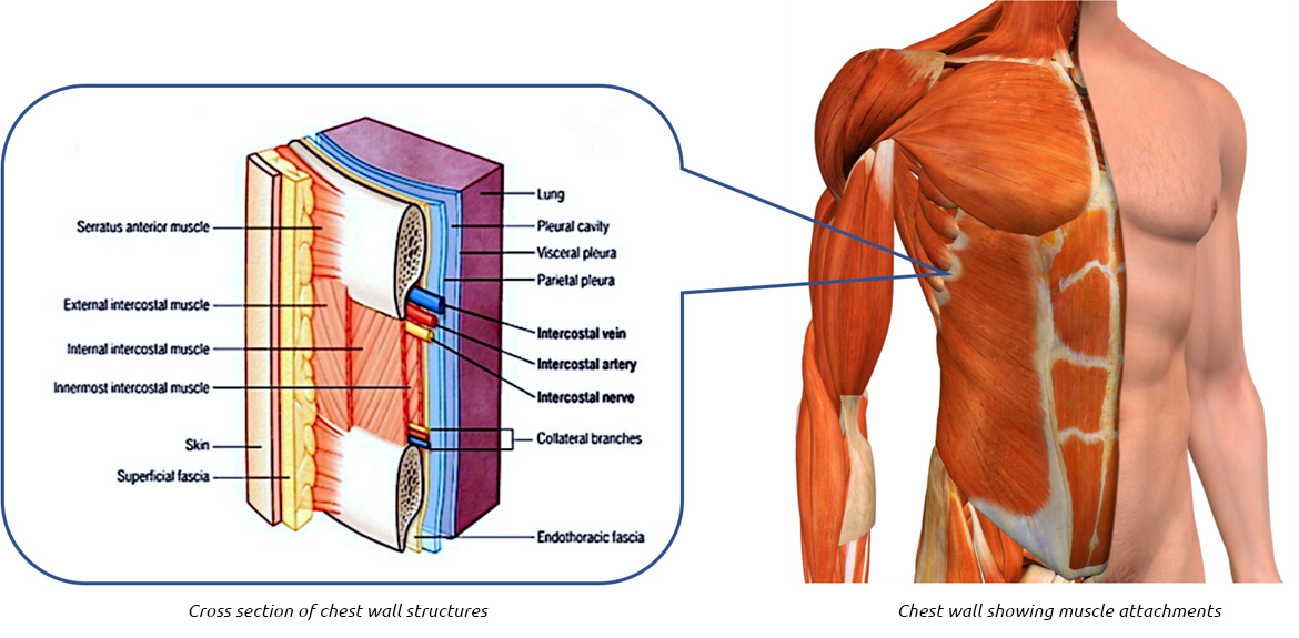
Fig. 3. Anterior chest wall showing the sternum. Note where the costal cartilages articulate with the sternum. In the intercostal space lie different structures: several kinds of intercostal muscles, intercostal arteries and associated veins, lymphatics, and nerves. (From Rendina EA, Ciccone AM. The intercostal space. Thorac Surg Clin 2007;17(4):491e501; with permission.) - "Relevant surgical anatomy of the chest wall."

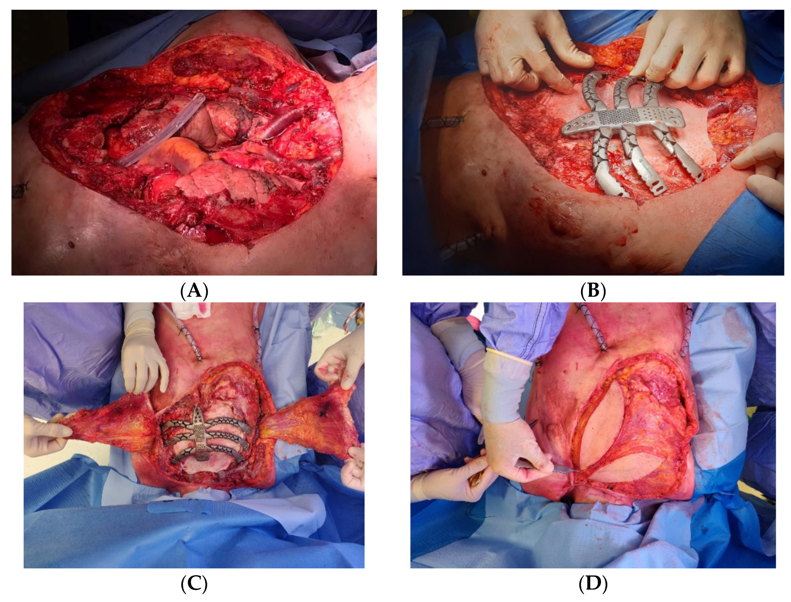
Chest Wall Resection

SURGICAL ANATOMY OF THE CHEST WALL
PERTINENT SURGICAL ANATOMY OF THE THORAX AND MEDIASTINUM

Chest Wall Anatomy: Overview, Gross Anatomy, Other Considerations

Mediastinum, Radiology Reference Article
Chest Wall Lumps Rib Injury Clinic

Illustration of the chest wall anatomy including suggested regional
Thorax Basicmedical Key
What Causes Empyema?
/jcm/jcm-11-05516/article_deploy/html/
Chest Wall: Anatomy Concise Medical Knowledge

Figure 1 from Relevant surgical anatomy of the chest wall.
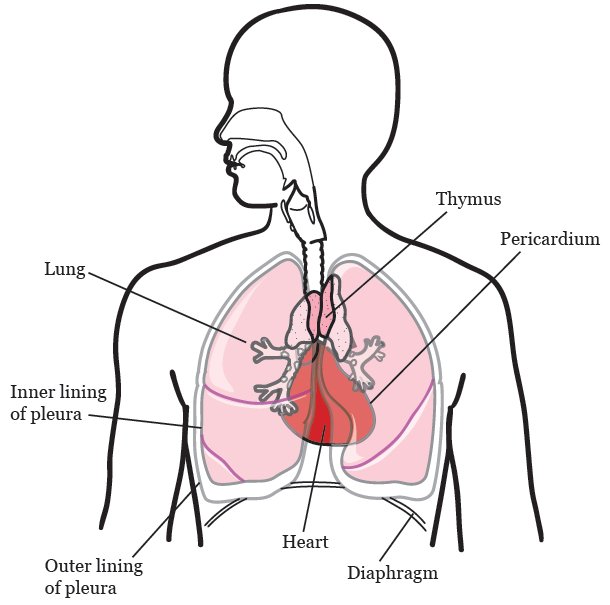
About Your Thoracic Surgery Memorial Sloan Kettering Cancer Center

SURGICAL ANATOMY OF THE CHEST WALL
Principles of Chest Wall Reconstruction
Anatomy of Chest with Drain in place – Medical Stock Images Company
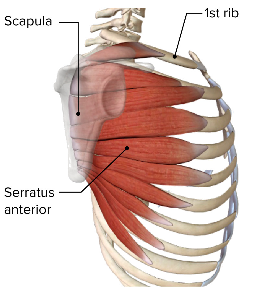
Muscles of the Thoracic Wall - Chest Muscles Anatomy
Chest Anatomy - wikiRadiography
Human chest anatomy Stock Photo by ©AnatomyInsider 129007478
Male chest anatomy of thorax with heart, veins, ar by StockTrek Images