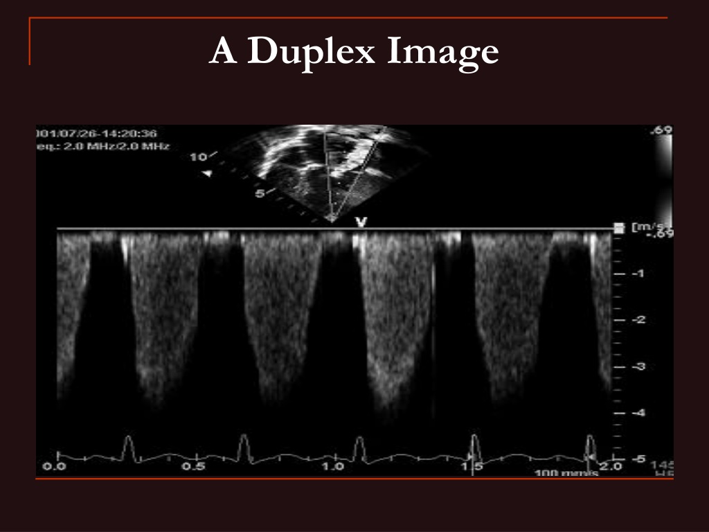
A duplex ultrasound output displaying the B-mode and colour flow
4.6 (133) In stock

PPT - Flow and Displacement Imaging PowerPoint Presentation, free download - ID:9689130
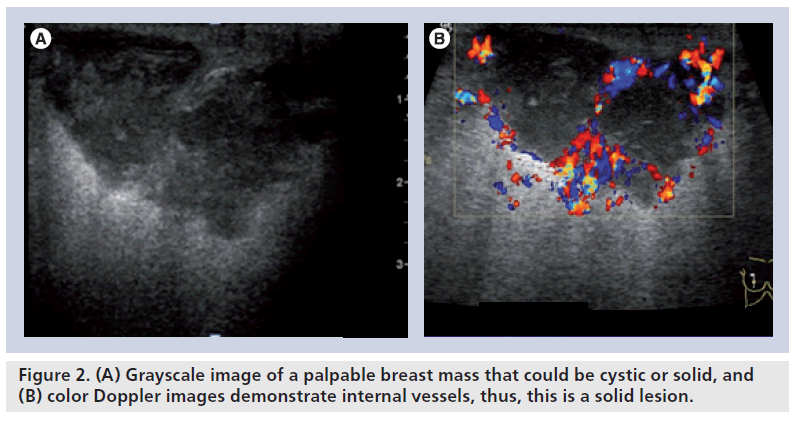
Color Doppler sonography: characterizing breast lesions

Cross-sectional B-mode duplex ultrasound scans of the right popliteal
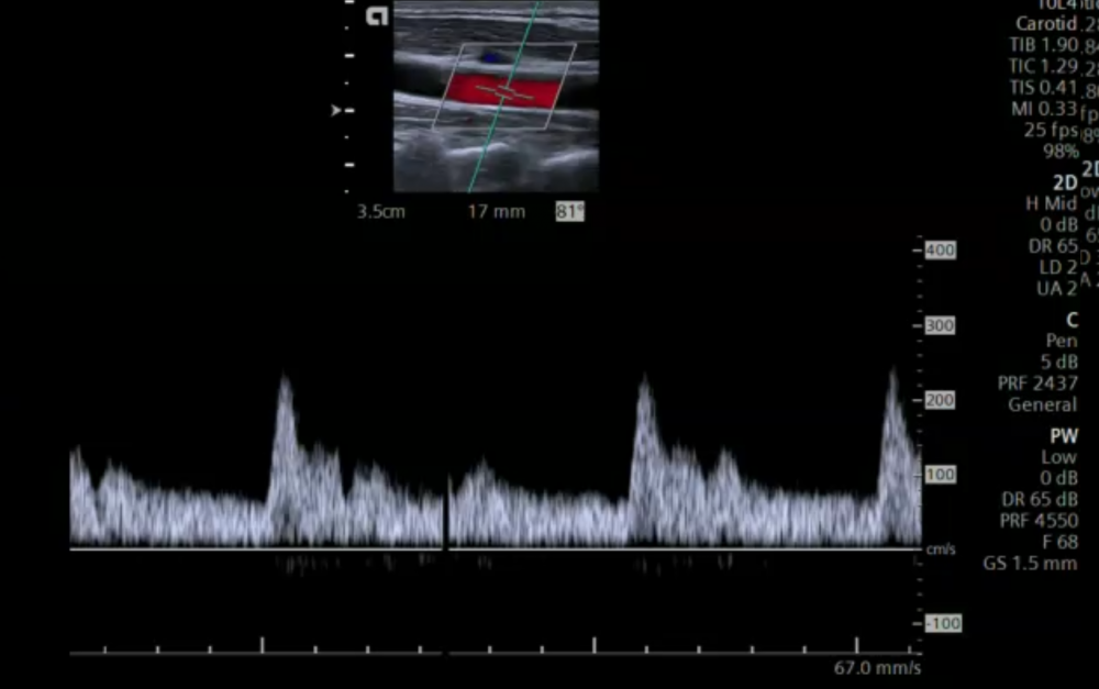
5. Color Doppler imaging of the carotid arteries

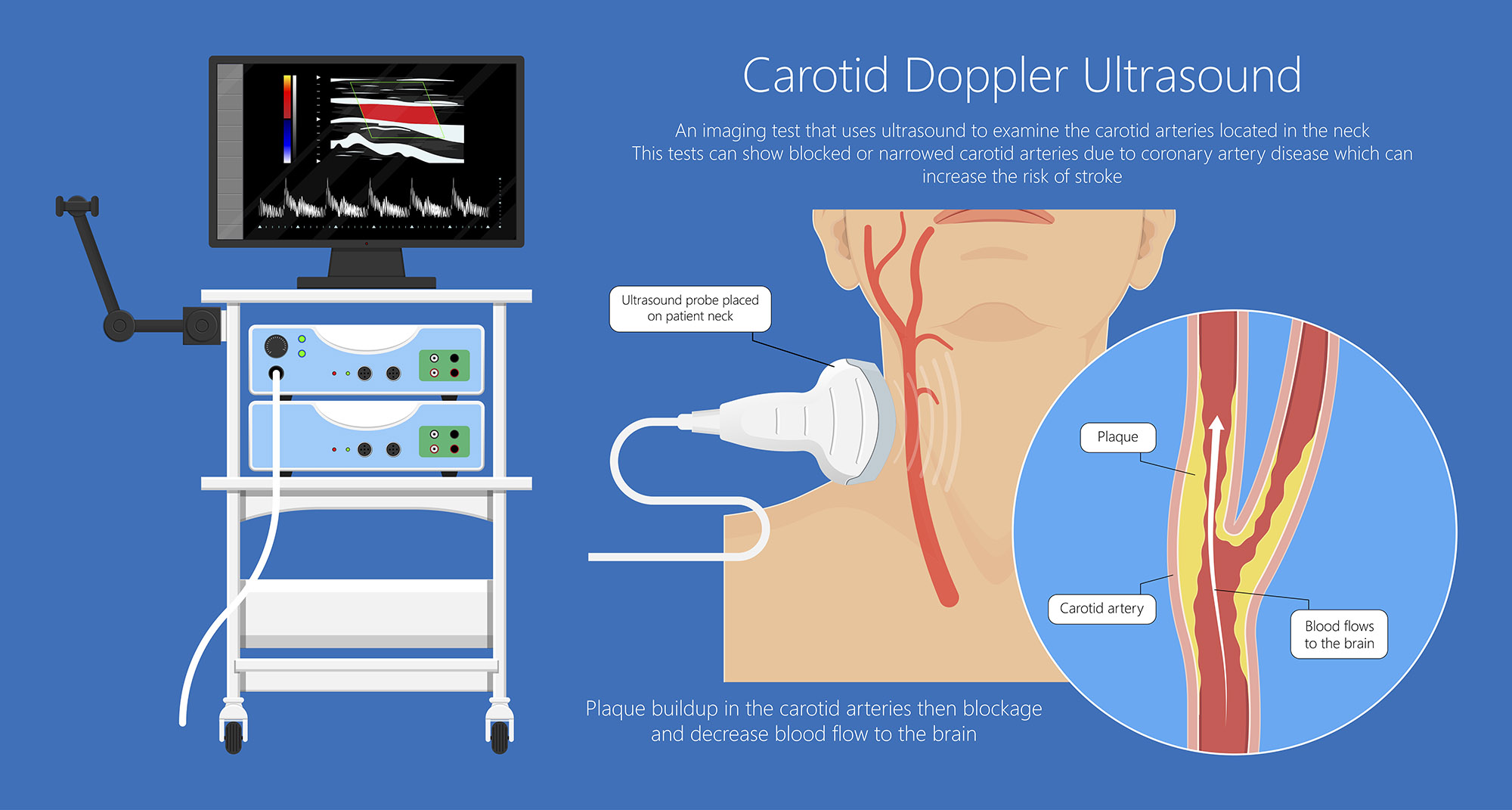
Imaging modalities to diagnose carotid artery stenosis: progress and prospect, BioMedical Engineering OnLine

PDF) Imaging modalities to diagnose carotid artery stenosis: Progress and prospect

Duplex ultrasound simulator examiner interface. (a) Color Doppler image

Our Protocol for Transabdominal Pelvic Vein Duplex Ultrasound - Endovascular Today

The use of colour Doppler imaging in the diagnosis of retinal detachment
Detect Vascular Disease

Application of B-flow imaging and its enhanced mode in perforator mapping - ScienceDirect

Schematic of Doppler ultrasound principle
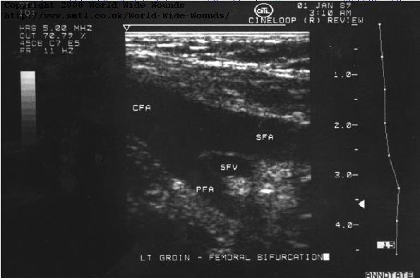
Review of duplex and colour Doppler imaging of lower-limb arteries and veins

PDF) Imaging modalities to diagnose carotid artery stenosis: Progress and prospect
B-Mode Ultrasound Innovations - Become a Sonographer
Ultrasound Machine Basics-Knobology, Probes, and Modes - POCUS 101
Modes Ultrasound A-mode- amplitude mode. B-mode- brightness mode. - ppt video online download
M-Mode Echocardiography and 2D Cardiac Measurements*
ACUSON Juniper is a High-Performance Shared Service Ultrasound
 FlexFit™ Drain Bulb Management Bra - Style No. B19
 NEW $98 Free People Movement You're A Peach Leggings Heather Gray
 Bali Women's Comfort Revolution EasyLite Shaping Wireless Bra
 UA Women's HeatGear Armour Compression Long Sleeve
 Contrast Lace Wireless Bra Comfy Breathable Front Buckle - Temu United Kingdom
 Buy White Thermal Wear for Women by JOCKEY Online