Figure 6 from Femoral Hernia: A Review of the Clinical Anatomy and
4.9 (565) In stock

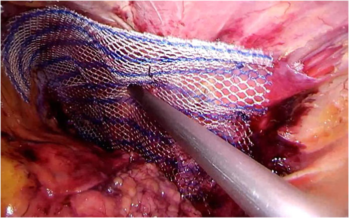
Figure 6. Femoral hernia repair in clean operation. (a) The narrow side of the mesh is sutured to Cooper’s ligament; (b) The mesh is sutured to the iliopubic tract or shelving portion of the inguinal ligament; (c) The posterior wall of the inguinal canal is reinforced, as in Lichtenstein’s repair. - "Femoral Hernia: A Review of the Clinical Anatomy and Surgical Treatment"

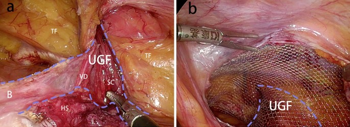
Figure 2 from Femoral Hernia: A Review of the Clinical Anatomy and Surgical Treatment

Femoral Hernia: A Review of the Clinical Anatomy and Surgical Treatment
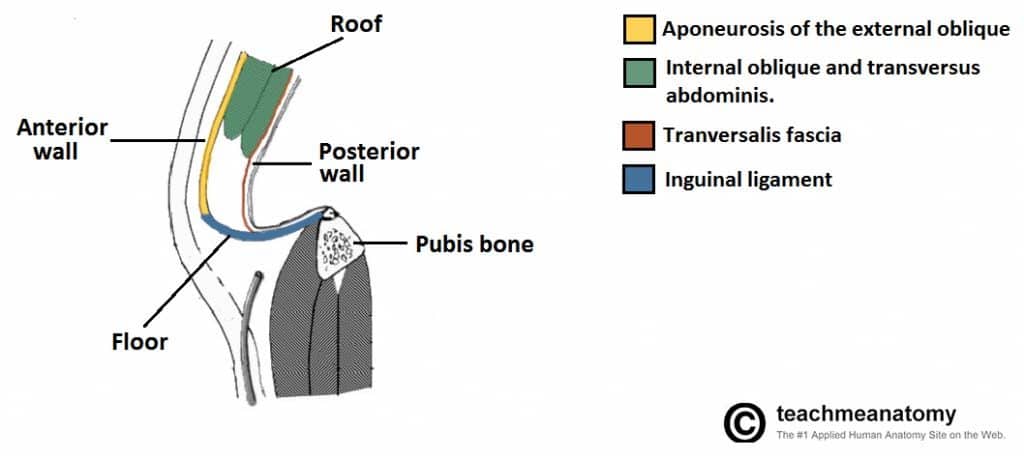
Femoral Hernia - Risk Factors - Clinical Features - Management - TeachMeSurgery
Femoral Hernia - A Review of Clinical Anatomy

PDF) Femoral Hernia: A Review of the Clinical Anatomy and Surgical Treatment
Surgical Techniques Development, Free Full-Text

Figure 6 from Femoral Hernia: A Review of the Clinical Anatomy and

The anatomical locations of the groin hernia defects. 1: Lateral
Femoral Hernia
Frontiers Publishing Partnerships Primary Lumbar Hernia, Review and Proposals for a Standardized Treatment
Embryonic developmental process and clinical anatomy of the preperitoneal fascia and its clinical significance
Inguinal Hernia - Classification - Management - TeachMeSurgery

Presence of the appedix vermiformis in the femoral hernia.

Femoral hernia

Femoral Hernia: A Review of the Clinical Anatomy and Surgical Treatment
Laparoscopic repair of an incarcerated femoral hernia - ScienceDirect
FEMORAL HERNIA REPAIR – OPEN – Dr. HH Pretorius
Femoral Hernia Repair, Dallas, TX
Femoral Hernia, Mr. Himaz Marzook
Laboratory value of patients before femoral hernia operation
 Vintage Laura Ashley Dress 1980s Strapless Cotton Turquoise Blue
- Empire' star Terrence Howard quitting his day job? 'I'm done with acting,' he says - Los Angeles Times
 Biustonosz Freya LOVELAND AA401002BOR Uw Plunge Bra Boudoir Noir Boudoir Noir
 Comfortable Seamless Adjustable Straps Bralettes Padded Bra
 Producto FAJA RELOJ DE ARENA CON CIERRE (VIENE REDUCIDA) de Fajas con Mary
 Winter Tweed Suits for Women