a B-mode image demonstrating a cervical length measurement. Cervical
5 (706) In stock

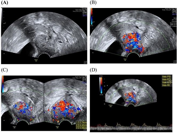
Download scientific diagram | a B-mode image demonstrating a cervical length measurement. Cervical tissue is outlined with the dotted line. The endocervical canal is demonstrated with a solid line. Two contiguous segments are often used when the cervix is not straight. The cervical length on this patient is 37.1 mm, which is in the normal range. b Ultrasound images illustrating the assessment of cervical consistency index (CCI). The left image is without pressure applied to the cervix. The right image is with pressure applied to the cervix by the transducer. CCI = 26 mm/32.9 mm × 100 = 79%. A smaller CCI is consistent with a softer cervix. c Strain elastography makes conclusions regarding tissue stiffness through observing deformations caused by probe pressure. Each color represents the difference in compressibility relative to the adjacent area. Softer tissue appears red while firmer tissue is assigned to blue from publication: Evolving cervical imaging technologies to predict preterm birth | Preterm birth, defined as delivery at less than 37 weeks’ gestation, increases maternal-fetal morbidity and mortality and places heavy financial and emotional burdens on families and society. Although premature cervical remodeling is a major factor in many preterm deliveries, | Preterm Birth, Elasticity Imaging Techniques and Elastography | ResearchGate, the professional network for scientists.
Measurement of cervical length using transvaginal sonography for
Significance of transvaginal sonographic assessment of cervical

Molly J. Stout's research works Concordia University–Ann Arbor

Sonographic assessment of cervical length and the risk of preterm

Sarah ENGLAND Washington University in St. Louis, Missouri

JCM, Free Full-Text

Cervical length measurement: Comparison of transabdominal and

Evolving cervical imaging technologies to predict preterm birth

Lihong V. Wang's research works

How to measure cervical length

a B-mode image demonstrating a cervical length measurement

a B-mode image demonstrating a cervical length measurement

Changes in cervical elastography, cervical length and endocervical

📃 Cervical incompetence, herniated amniotic sac through the
Updated applications of Ultrasound in Uterine Cervical Cancer
File:Dilated cardiomyopathy B-Mode.jpg - Wikipedia
Ultrasound Physics and Technical Facts for the Beginner
 Seward Round Braided Rug
- Kriss Soonik - Silvia Fishnet Bra Maike Fishnet Knickers www.kriss
 knix, Intimates & Sleepwear, Knix Shadow Mesh Tshirt Bra Size Xxxxl Orchid
 Chicago Bulls launch Ring of Honor, announce inaugural class
 Men's Underwear with A Secret Front Stash Pocket Panties, Small
 Teen Period Underwear - Bikini, Nude
