A novel approach to sonographic examination in a patient with a
4.8 (335) In stock
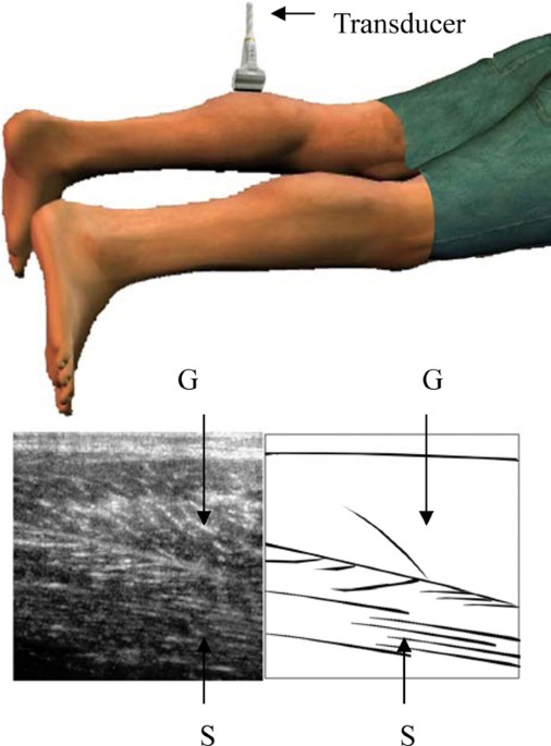
Introduction Rupture of the distal musculotendinous junction of the medial head of the gastrocnemius, also known as "tennis leg", can be readily examined using a soft tissue ultrasound. Loss of muscle fiber continuity and the occurrence of bloody fluid accumulation can be observed using ultrasound with the patient in the prone position; however, some cases may have normal ultrasound findings in this conventional position. We report a case of a middle-aged man with tennis leg. Ultrasound examination had normal findings during the first two attempts. During the third attempt, with the patient's calf muscles examined in an unconventional knee flexed position, sonographic findings resembling tennis leg were detected. Case presentation A 60-year-old man in good health visited our rehabilitation clinic complaining of left calf muscle pain. On suspicion of a ruptured left medial head gastrocnemius muscle, a soft tissue ultrasound examination was performed. An ultrasound examination revealed symmetrical findings of bilateral calf muscles without evidence of muscle rupture. A roentgenogram of the left lower limb did not reveal any bony lesions. An ultrasound examination one week later also revealed negative sonographic findings. However, he still complained of persistent pain in his left calf area. A different ultrasound examination approach was then performed with the patient lying in the supine position with his knee flexed at 90 degrees. The transducer was then placed pointing upwards to examine the muscles and well-defined anechoic fluid collections with areas of hypoechoic surroundings were observed. Conclusion For patients suffering from calf muscle area pain and suspicion of tennis leg, a soft tissue ultrasound is a simple tool to confirm the diagnosis. However, in the case of negative sonographic findings, we recommend trying a different positional approach to examine the calf muscles by ultrasound before the diagnosis of tennis leg can be ruled out.

Whole Body Ultrasonography in the Critically Ill

PDF) A Nearly Missed Pancoast Tumour From Isolated Persistent Leg Pain

Ultrasound: A Practical Approach to Clinical Problems

Ultrasound imaging of the axilla, Insights into Imaging

Ultrasound Exam Approach in Trauma Patients

Revolutionising Ultrasound: Advancements in Hand Imaging with

Ultrasound Exam Approach in Trauma Patients
Frontiers Application of ultrasound in cardiovascular

Free Ultrasound e-Books - Ultrasound Training

PDF) A Novel Method of Laparoscopic Approach in a Giant Bladder

Gynecology/Pelvic Ultrasound Made Easy: Step-By-Step Guide - POCUS 101
PDF) The Role of Ultrasound and Shear-Wave Elastography in

Examination Review for Ultrasound: SPI: Sonographic Principles
Calf Strains - Holistic Bodyworks
Calf Pain: Causes, Treatment, and When to See a Healthcare Provider
Calf Strain - Lower Leg - Conditions - Musculoskeletal - What We
 Rosegal Women Plus Size Bell Bottoms Gothic Flare
 Prettylittlething Blue Logo Short Sleeved Bodysuit
 Skinfood Forest Dining Bare Foundation 01 Natural Beige (35g), Beauty & Personal Care, Face, Makeup on Carousell
 ElenaDressy Vestidos Largos de Noche de satén, línea A, Sencillos
 Ally Bra - Vitesse Athletics
 42C Size Bra - Buy 42C Level 3 Bra Online
 Just My Size® Women's Hipster 5 Pack Tagless & Plus Size & Smooth Stretch NEW
 sexy chick nude ready for cock - hot amateur brunette showing C cup tits Porno Photo - EPORNER
 Пряжа Pura Lana Italia LINO Italiano 650 м - Магазин Анже
 RBX Activewear Scuba 1/2 Zip Sweatshirt for Women, Lightweight
 Faja Moldeadora Cuerpo Adelgazante Hombre Reducir Cintura - Temu